Authors
Bryttin Boyde & Hannah Selvey
Abstract
Studies have shown that carnivores in captivity that are fed soft diets have more periodontal problems and that the morphology of their skeletons, skulls specifically, responds to environmental and behavior factors. Considering that captive diet affects teeth and gums, we have conducted research on Dr. Adam Hartstone-Rose’s hypothesis that variation will also be discernible through the examination of cranial morphology. We compared captive and wild lions Panthera leo and tigers Panthera tigris to see if differences were present, then analyzed data collected by a microscribe using a 3D morphometric analysis program. Next, Principal Component Analysis was completed to account for variation across eighty-one specimens, each with forty-three comparable landmarks. We found that captivity status is evident in felid cranial morphology and is even more pronounced than features of sexual dimorphism. The implications of this research could serve as possible bases for the reformation of captive diet. Thus, if a correlation between mechanical diet and detrimental effects on captive felids can be shown through research, measures to fix these may be taken.
Introduction
Captive carnivores experience health disparities due to inadequate diets (Haberstroh
et al. 1984). The genus Panthera (“big cats”) is a prime example of a carnivoran group
that, when residing in captivity, is morphologically impacted by environmental factors
such as mechanical diet. Although zoos and rescue centers attempt to provide food
that mimics the nutrition these animals would acquire in the wild (Glatt et al. 2008),
nutrition is not the only criteria a captive diet must meet in order to fully satisfy
a wild animal’s physiological and psychological requirements: the consistency and
texture of an animal’s diet, particularly those lacking the mechanical properties
of a wild diet, such as bones and other elements that require chewing, can have adverse
long-term effects on zoo animals, specifically carnivores (Glatt et al. 2008)
Enrichment beyond nutrition is important for zoo animals and much evidence supports
the claim that captive animals’ diet affects them adversely. Skibiel et al. (2007)
considered the lack of stimulation captive felids experience as a result of the absence
of predator-prey relationships in their environments. Their study was based on the
premise that captive animals are under-stimulated, both cognitively and physically,
which creates negative repercussions on the animals that is then evident in their
behavior. The animals’ temperaments and overall health were tested by supplementing
their normal (mechanically and psychologically unstimulating) diets with bones and
frozen fish. Results of this study thus confirmed that a diet closer to that which
would be found in nature could possibly prevent negative changes due to under stimulation,
both psychological and physiological, in captive animals.
Research has shown that, in addition to being under-stimulated, certain captive carnivores
have experienced visible health disparities due to inadequate diets (Haberstroh et
al. 1984). Captive Amur tigers, (P tigris altaica) have experienced oral health problems
that researchers attribute to behavioral and environmental factors, including being
fed soft, commercial, meat-based diets (Haberstroh et al. 1984). Haberstroh and colleagues
suggests that factors beyond genetics influence dental pathologies. Despite a sufficient
amount of nutrients, carnivores in zoos typically have more dental problems than those
in the wild due to the lack of abrasive action that usually accompanies chewing on
bones (Haberstroh et al. 1984). A few documented deformities include periodontal disease
(Haberstroh et al. 1984), plaque buildup (Glatt et al. 2008) and hypoplasia (O’Regan
et al. 2005).
Indirect “domestication” of animals exists and is impacting their morphology and creating
further occurrences of dental pathologies. O’Regan et al. (2005) describes this phenomenon,
discussing captivity’s unintended consequences, such as preventing the preservation
of animals as they exist in the wild. Although recent measures to ameliorate the negative
consequences of inadequate mechanical diets are currently under review, North American
collections mostly still feed their captive felines a diet with physical properties
that hardly require chewing to digest (O’Regan et al. 2005). Zoological morphologists
have confirmed that differences in skull shape, including cranial thickness in captive
lions (P. leo) have also been severely impacted by captive diets (O’Regan et al. 2005).
Hartstone-Rose et al. (2012) found support for the idea that what carnivores eat will
ultimately affect their muscular and osteological masticatory architecture. Through
analysis of masticatory muscles in captive felines, this research confirmed that a
stronger bite force is correlated directly to the amount and size of obdurate, or
hard, foods that the animals have consumed, thus supporting the notion that geometrical
properties of food are indeed correlated to the feeding architecture of the studied
carnivorous consumers (Hartstone-Rose et al. 2012). An additional study concerns reptilian
consumers of obdurate foods and makes connections between durophagy, the practice
of eating hard foods, and head shape (Schaerlaeken et al. 2012). Durophagous lizards
were found to have significantly different head shapes and sizes from lizards that
preyed on softer organisms. This study’s findings support the notion that studying
resource use can aid in making deductions about a carnivore’s functional capacity.
Because specialized diets have been correlated with the mechanisms required for digestion
in carnivorous lizards, we can expect to find similar correlations between carnivorous
felids and their diets in captivity and in the wild. A study conducted by Hartstone-Rose
et al. (2013) supported that studying morphology of durophagous carnivores can tell
us about diet. Using Principal Component Analysis (PCA), we can explore the morphological
similarities and differences across a population of mixed captivity statuses and make
inferences about the impact of captivity based on our observations.
One important factor driving the differences within this population is likely to be
sex. Sexual dimorphism is evident in both lions and tigers (Mazak 2004 & Naples et
al. 2012). At times, the sexes of tigers can even cause them to appear to be two difference
species; in fact, an experiment using PCA yielded results showing that sex accounted
for over seventy-seven percent of intraspecific variance (Mazak 2004). According to
Mazak (2004), a large portion of the factors influencing the differences across sexes
is related to morphology that is strongly correlated with predatory function, such
as the rostra and zygomatic arches. This is significant when considering that lions
are the most sexually dimorphic of the big cats (Naples et al.2012). A study of male
and female lion specimens yielded results with substantial differences in the porosity
of the skulls of the males as opposed to the females, which the authors deduced to
be correlated with the lifestyle behavior of the felids (Naples et al.2012).
If indeed morphology is related to diet, we hypothesize that differences in mechanical
diet across captive and wild populations of lions and tigers will be statistically
discernible through PCA and with three-dimensional examination of the felids’ skulls.
With knowledge of the diet of our carnivoran population, we will work backwards to
see how morphology is specifically affected by that diet and eventually discover the
ideal provisions for captive felids. We expect that, in addition to species, sex and
captivity status will be responsible for the differences between the individuals in
the sample population. Studying the impact of captivity on carnivores is vital to
ensure the future health and safety of captive animals (Haberstroh et al. 1984). Therefore,
in order to provide the best possible environments for carnivores in captivity, we
must understand the impact captivity has on those carnivores.
Methods
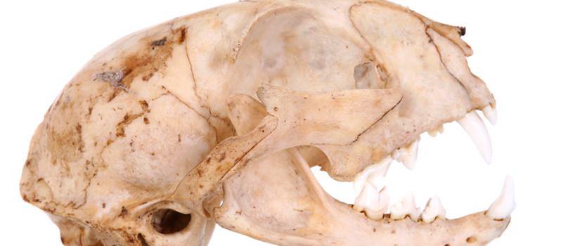
To determine if differences exist across the two captivity statuses, captive and wild,
of large felids, we implemented statistical analysis on three-dimensional renderings
of the sample population of lion and tiger specimens (See Table 1). The sample population
of specimens was sorted according to species, captivity status, and sex.
For the sake of this paper, “wild” refers to mature individuals who did not live in
captivity and “captive” refers to mature individuals who resided in zoos or rescues.
Samples were obtained from collections at the American Museum of Natural History (AMNH)
the Smithsonial (USNM) and the research collection of Dr. Hartstone-Rose (University
of South Carolina School of Medicine). These specimens originated at the Bronx Zoo,
Central Park Zoo, New York Zoo, New York Zoo Society, New York Zoo Gardens, New York
Park Commission, National Zoological Park (Smithsonian), Toledo Zoological Society,
Academy of Natural Science, Barnum and Bailey, Prospect Park Zoo, and the Carolina
Tiger Rescue.
Forty-three landmarks on the skulls of each specimen were recorded with a microscribe
(Solution Technologies, Inc,). Microscribes can record common landmarks across objects,
such as skulls, providing the shape of those objects relative to each other, using
(x, y, and z) coordinates. Coordinates are used to code for three-dimensional renderings,
which can then be entered into a Microsoft Excel document and viewed with various
computer programs. Forty-three landmarks are as follows: 1 Foramen Magnum inferior,
2 Foramen Magnum superior, 3 Inion, 4 Vertex, 5 Nasion, 6 Rhinion, 7 Alveolare, 8
Infradentale, 9 Antero-lateral nasal corner L, 10 Buccal edge of maxilla at Canine
L, 11 Distal P4* L, 12 Orbitale L, 13 Lateral orbit L, 14 Superior orbit L, 15 Medial
orbit L, 16 Coronion (Coronoid tip )L, 17 Zygion L, 18 Porion L, 19 Tip of mandibular
angle L, 20Antero-lateral nasal corner R, 21 Buccal edge of maxilla at Canine R, 22
Distal P4 R, 23 Orbitale R, 24 Lateral orbit R, 25 Superior orbit L, 26 Medial orbit
L, 27 Coronion (Coronoid tip )R, 28 Zygion R, 29 Porion R, 30 Tip of mandibular angle
R, 31 Anterior edge of Canine at premax/max sutures L, 32 Posterior edge of Canine
L, 33 Anterior edge of lower p3 L, 34 Anterior edge of P4 L, 35 Anterior edge of masseter
origin L, 36 Posterior edge of masseter origin L, 37 Superior edge of zygomatic arch
at suture L, 38 Superior edge of masseter origin at thickest L, 39 Inferior edge of
masseter origin at thickest L, 40 Anterio-superior corner of temporalis origin L,
41 Posterio-superior corner of temporalis origin L 42 Posterio-inferior corner of
temporalis origin L, 43 Anterior-inferior corner of temporalis origin L.
The landmarks for each specimen were entered into an Excel Spreadsheet with columns
respectively labeled: x, y, and z. The points for each specimen were entered into
a text-only document, which we coded for upload into the three-dimensional geometric
morphometric analysis program, Morphologika, (version 2.5). Morphologika translates
the landmarks as recorded by the microscribe and plots them into an object that can
then be viewed, moved, and analyzed (See wireframes in Figure 1 & Figure 2).
Renderings for each specimen were uploaded into Morphologika and we used Procrustes
analysis to superimpose the landmarks across specimens so that they could be compared
to each other in aligned space (O’Higgins and Jones 2006). Procrustes analysis accounts
for changes that exist in coordinates due to the relative space where the specimen
was located during the data collection. Procrustes analysis aligns object by “minimizing
the sum of the squared distances between corresponding landmarks” (Von Cramon-Taubadel
2007). This prepares the three-dimensional population for Principal Component Analysis
(PCA).
We then ran a PCA in Morphologika, comparing all the specimens to determine what the
principle driving components were that accounted for the differences across specimens.
The PCA output was graphed in Morphologika after all the principle components were
calculated. We plotted PC1 and PC2 against each other (Figure 1), and PC2 and PC3
against each other on graphs (Figure 2). Because PCA only accounts for quantitative
data, we examined the graphs as well as the differences in the wireframes of the specimens,
generated by connecting some of the key landmarks, across the x-axis and y-axis to
determine the principle components, or strongest factors driving variation across
the sample population. Interpreting the markers for each type of specimen, we could
easily interpret the driving factors behind the first three principle components (See
Figure 1 & Figure 2).
Figure 1: The first two principle components of the lion and tiger 3D data. Individuals in the sample population are represented as they vary according to the two components. A clear distinction between species is visible along the x-axis and a clear distinction between captivity statuses is visible on the y-axis. Morphological variation is (marked) at each extreme. The right and left extremes of the x-axis represent P. leo and P. tigris specimens respectively. The upper range of the y-axis represents morphology characteristic of the captive felids and the lower range represents wild. A) Differences in rostrum length as it varies across (1) P. tigris and (2) P. leo. B) Variance of mandibular angle across (1) P. tigris, and (2) P. leo. C) Biangular-anterior mandibular angle, or dome shape is described in (1) captive and (2) wild specimens. D) Skull width across (1) captive and (2) wild specimens.
Figure 2: The second and third principle components of the lion and tiger 3D data. Individuals in the sample population are represented as they vary according to the two components. A clear distinction between captivity statuses is visible along the x-axis and a clear distinction between sexes is visible on the y-axis. Morphological variation is (marked) at each extreme. The right and left extremes of the x-axis represent captive and wild specimens respectively. The upper range of the y-axis represents morphology characteristic of males and the lower range represents females. A) Differences in the shape of the maxillary area as it varies across (1) males and (2) females. B) Variance of mandibular angle across (1) males and (2) females.
Table 2: PCA output from Morphologika 2.5 with forty-three points of comparison: eighty factors influencing variance across population were found. The Eigen value, percentage of total variance, and cumulative variance are described in this table.
Results
Graphing the PCA showed that species was the first principle component, accounting
for 21.28% of the variation, and visibly separating the groups into lions and tigers
with almost zero overlap on the x-axis (See Figure 1 & Table 2). The second principle
component clearly represented captivity status, accounting for 15.58% of the variance,
and separating the wild and captive specimens across the y-axis. The third principle
component represented the sex of the individuals, accounting for 7.97% of the variance.
Figure 2 shows the second principle component, captivity status, plotted against the
third, with females occupying mostly the lower extreme of the y-axis and males occupying
the higher extreme.
Differences in morphology were evident at each extreme of the x-axis and y-axis of
each PCA test (See Fig. 1 & 2). Among some of the differences were the length of the
rostrum, mandibular angle, flexion of the mandibular angles relative to the mandibular
symphysis, and the width of the skull. Rostral length differed across species, as
tigers were shown to have shorter rostra than lions, which is a trait that has been
described by Sunquist (2002) and Christiansen (2007). Christiansen also describes
increased nasal height in tigers and differentiates between canine heights across
lions and tigers. Mandibular angles also varied across the species, as tigers showed
mandibles wider at the top (i.e., bi-coronal breadth) and lions showed the widest
point at the base of the mandible (i.e., bi-angular breadth).
Different skull shapes and differences in width were also observable across captivity
status. Mandibular angle and rostrum length varied across lion and tiger individuals
(Figure 1). Captive individuals appeared to have flatter heads than the wild specimens
against whom they were compared. The width of the skull across specimens was also
noticeably different, as captives seemed to have wider skulls than wilds. Within both
species, mandibular angle variation was also evident across males and females.
Discussion
As expected, the first driving factor, PC1 (21.28%) separated the two species, as
obvious phenotypic differences exist between tiger and lion individuals (Sunquist
and Sunquist 2002). As you move further right on the x-axis of the graph shown on
Figure 1, you can see that the frequency of tiger individuals declines and the frequency
of lion individuals increase. Unexpectedly, the second most important source of variation
(PC2) was most influenced by captive status (15.57%) and not sex (which emerged as
the key factor in PC3). This means that, after species, captive status is the most
discernable characteristic across this population. Previous studies on both captive
tiger and lion individuals have yielded results supporting the idea that captivity
status affects morphology. Geordie Duckler attributed malformations in the external
occipital region of tiger skulls to phenotypic plasticity, judging that significant
differences between the examined captive and wild specimens of the study were due
to “reduced jaw activity” (Duckler 1998). A similar observation was made by O’Regan,
examining the cranial thickness in captive lion individuals (O’Regan 2005).
Differences across species were evident upon examination of the wireframe renderings
of the specimens in Morphologika. One difference is a shortened rostrum in the tiger
specimens relative to the lion specimens, which is consistent with descriptions of
tigers (Sunquist and Sunquist 2002). This is a possible topic for future research.
Mandibular angles also varied across the species, as tigers exhibited wider bi-coronal
breadths and lions exhibited wider bi-angular breadth. The results of the PCA output
are encouraging, demonstrating the ability of statistical analysis to account for
observable qualitative differences across specimens. Variation of skull width and
in biangular-anterior mandibular angle, or dome shape was observed differences across
captivity status. Wild specimens were found to have more robust domes, than captive
(Figure 1). Relatedly, captive specimens had greater skull widths relative to length
than wild.
The third principle component, observed to be sex, accounted for nearly eight percent
of the variance across the sample population (7.97%, respectively.) The fact that
sex was the third principle component, behind species and captivity status, suggests
that it is easier to tell the difference between captive and wild felids than it is
to tell the difference between the sexes of the two species. One explanation for this
phenomenon is that behavioral differences between sexes that occur in the wild due
to hunting do not occur in captivity and therefore do not contribute to sexual dimorphism
in captivity. A further question comes up in the analysis of these data: are females
and males affected by captivity to different extents?
The results of this study supported the captivity status hypothesis and statistically
confirmed that observable differences in cranial morphology do exist across species.
These results are especially significant because sexual dimorphism is a known characteristic
of both lion and tiger species (Naples 2012, Mazaak 2004). Whether the differences
that occur are actually due to mechanical diet is a persistent question. The strength
of captivity status as a component of difference in the data also opens up a new line
of inquiry about sexual dimorphism. There are two questions to consider: How much
do the mechanical properties of food really affect felids? In what other ways captivity
status affects morphology of captive felids?
A study to further investigate captivity’s impact on cranial morphology will include
more species of large carnivores as well as a control group. A type of zoo animal
such as Zalophus californicus, the California sea lion, with a diet primarily made
up of fish in captivity and the wild should therefore exhibit little to no differences
in cranial morphology across captivity status if the morphology is mostly influenced
by the food’s mechanical properties. If there are differences in morphology across
captivity status, regardless of diet, other possible factors contributing to the differences
in cranial morphology across captivity statuses may include genetic issues, for example
those stemming from inbreeding.
If further studies yield similar results to those of this analysis and continue to
show a direct relationship between captive diet and changes in cranial morphology
of carnivores, these studies could be a possible basis for handlers and animal dietitians
to rethink their policies regarding the mechanical diets of captive carnivores. If
a mechanical diet that requires further engagement of masticatory muscles, because
nutrients alone are insufficient for the proper maturation and health of captive felids,
zoos must take proper measures to assure the health of captive specimens.
Acknowledgements
Thanks to Dr. Adam Hartstone-Rose for giving us this study to work on and guiding the research process. Thanks to Dr. Erin Connolly for reviewing this paper and providing guidance in the approach to writing it. Thanks to Joseph Villari and Kristen MacNeill for collecting the data used in this study. Thanks to “Elvin” Boone for technical support and helping with the installation of Morphologika at the workstation.
About the Authors
 Bryttin Boyde
My name is Bryttin Boyde and I am from The Woodlands, Tx. I am currently a junior at USC and in the spring of 2015 I will graduate with a degree in Anthropology and minors in Biology and Chemistry. As a member of the Honors College I have experienced the opportunity of being awarded a SURF grant in order to pursue biological anthropology research in Dr. Adam Hartstone-Rose’s comparative anatomy lab at the USC SOM and have grown so much as a student researcher. The motivation behind this project comes from my desire to expand my knowledge of anatomy at the osteological level in order to fully comprehend everything that goes into masticatory muscle architecture, motor development and the effects of diet on both animals and humans. Being able to assist in this research has impacted me by directly enhancing my understanding of morphometrics and the effects that diet has on both captive and wild animals, and also how much attention this issue deserves. Moreover, the process of generating a scholarly research article has confirmed in me my want to pursue further research and publish in a well-respected anthropology journal. Additionally, the opportunity to publish in Caravel will add merit and accomplishment to my resume and help shape me as a skilled and experienced applicant to both national fellowships and graduate schools. My future plans include attending graduate school to get a Ph.D. in biological anthropology with emphasis on comparative anatomy, osteology and functional morphology with the end goal of being a researcher and teaching at the university level. All of this would not be possible without the assistance and support of my mentor, Dr. Hartstone-Rose, my research partner, Hannah Selvey and the Honors College SURF grant.
 Hannah Selvey
In 2015, I will graduate with a degree in biology and anthropology and continue to a graduate program in evolutionary biology with an emphasis on comparative anatomy. Acceptance into the TRiO Ronald E. McNair Postbaccalaureate Achievement Program taught me how to conduct scholarly research and encouraged me to continue to a Ph.D. level in pursuit of learning about and teaching and biological anthropology. The program also enabled me to apply for Magellan Guarantee funding, with which I have been continuing my research. I have also taken advantage of the undergraduate research funding opportunities in the Honors College to continue my work with my mentor, Dr. Hartstone-Rose, with the Exploration Grant this spring. My first paper will be in review for publication in a scholarly journal by the spring. Initially, Dr. Hartstone-Rose helped me pick this topic, due to my interest in cranial comparative anatomy, and has since helped me develop it into the paper that it is today. The most memorable part of my research process thus far has been probably collecting morphometric data back in my hometown, at the American Museum of Natural History. This research is particularly exciting because it has shown me that I really do want to take the research route to a professional career in what I love to learn about plan to eventually teach. In addition to the personal realizations and new goals I now have as a result of conducting research as an undergraduate, I now also have experience presenting my research, as I conducted oral presentations at the 2013 SC McNair Research Symposium and the 2013 SAEOPP McNair/SSS Scholars Research Conference, thanks to Melissa Kupfer with TRiO at USC, Dr. Hartstone-Rose, and Joe Valleri who helped with data collection.
References
Christiansen, P. 2007. Canine morphology in the larger Felidae: implications for feeding ecology. Biological Journal of the Linnean Societ 91: 573-592.
Duckler, G. L. 1997. Previously Undescribed Features in the Temporalis and Masseteric Musculature of Several Large Felids Raised in Captivity. Zoo Biology 16:187-191.
Duckler, G. L. 1998. An Unusual Osteological Formation in the Posterior Skulls of Captive Tigers (Panthera tigris) Zoo Biology 17:135-142.
Glatt, S. E., K. E. Francl, and J. L. Scheels 2008. A Survey of Current Dental Problems and Treatments of Zoo Animals. International Zoo Yearbook 42(1): 206–213.
Haberstroh, L. I., D. E. Ullrey, J. G. Sikarski, et al. 1984. Diet and Oral Health in Captive Amur Tigers (Panthera tigris altaica). The Journal of Zoo Animal Medicine 15(4): 142–146.
Hartstone-Rose, A J. M. G. Perry, and C. J. Morrow 2012. Bite Force Estimation and the Fiber Architecture of Felid Masticatory Muscles. The Anatomical Record: Advances in Integrative Anatomy and Evolutionary Biology 295(8): 1336–1351.
Hartstone-Rose, A, Stynder, DD 2013. Hypercarnivory, durophagy or generalized carnivory in the Mio-Pliocene hyaenids of South Africa? South African Journal of Science 109(5-6).
Mazak, J. H. On the Sexual Dimorphism in the Skull of the Tiger (Panthera tigris) 2004. Mammalian Biology 69(6): 392-400.
Naples, V. L., and B. M. Rothschild 2012. Sex Determination in Lions (Panthera leo, Felidae): a Novel Method of Distinguishing Male and Female Skulls. Mammalia 76(1).
O’Higgins P and N. Jones (1998) Facial growth in Cercocebus torquatus: An application of three dimensional geometric morphometric techniques to the study of morphological variation. Journal of Anatomy. 193: 251-272
O’Regan, H. J., and A. C. Kitchener 2005. The Effects of Captivity on the Morphology of Captive, Domesticated and Feral Mammals. Mammal Review 35(3-4): 215–230.
Schaerlaeken, V., V. Holanova, R. Boistel, et al. 2012. Built to Bite: Feeding Kinematics, Bite Forces, and Head Shape of a Specialized Durophagous Lizard, Dracaena Guianensis (Teiidae). Journal of Experimental Zoology Part A: Ecological Genetics and Physiology 317(6): 371–381.
Skibiel, A. L., H. S. Trevino, and K. Naugher 2007. Comparison of Several Types of Enrichment for Captive Felids. Zoo Biology 26(5): 371–381.
Sunquist, M. E., & Sunquist, F. (2002). Wild Cats of the World. Chicago: The University of Chicago Press.
Von Cramon-Taubadel, N., B. C. Frazier, and M. Mirazón Lahr 2007. The Problem of Assessing Landmark Error in Geometric Morphometrics: Theory, Methods, and Modifications. American Journal of Physical Anthropology 134(1): 24–35.
Software/Analysis Programs
”Introduction to Microscribe and Morphologika”
2011 Harcourt-Smith, Will. 35min. Paleo-Tech Concepts, Inc. Youtube.
Microsoft (2010) Microsoft Excel [computer software]. Redmond, Washington: Microsoft.
O’Higgins, P, Jones, N 2006. Morphologika (version 2.5) [computer software]. Hull York Medical School. http://www.york.ac.uk/res/fme/resources/software.html